Brush Biopsy: Revolutionizing Oral Cancer Detection in Toronto
Your regular dental visit may soon include a swift and painless screening for oral cancer, a breakthrough technology emerging from research at the University of Illinois Chicago. This article discusses how the innovative brush biopsy technique could transform the landscape of oral cancer diagnosis within dental practices in Toronto and the Greater Toronto Area (GTA).
Understanding Oral Cancer
Oral cancer refers to malignancies that develop in various parts of the mouth, including the lips, tongue, gums, under the tongue, and mouth roof. These cancers can be life-threatening, particularly if not caught early, which makes regular screening a critical preventive tool for both patients and providers.
Types of Oral Cancer
- Squamous Cell Carcinoma: The most prevalent type, originating from the flat epithelial cells lining the interior of the oral cavity.
Risk Factors
- Tobacco use (smoking or chewing)
- Heavy consumption of alcohol
- HPV infection, especially HPV-16
- Prolonged sun exposure (particularly for lip cancers)
- Poor oral hygiene and nutritional deficiencies
- A family history of oral cancers
Symptoms
Initially, oral cancer can present with subtle signs. Patients should be aware of persistent sores, red or white patches, unexplained bleeding or pain, difficulty swallowing, oral numbness, lumps, or a change in voice that doesn’t resolve.
Importance of Early Detection

Detecting oral cancer in its early stages significantly improves prognosis. Non-invasive technologies, including the VELScope, available through EBIKO Dental, allow clinics in Toronto to offer fast, real-time detection of abnormal mucosal tissues—reducing diagnostic delay and improving patient outcomes.
Diagnosis and Treatment
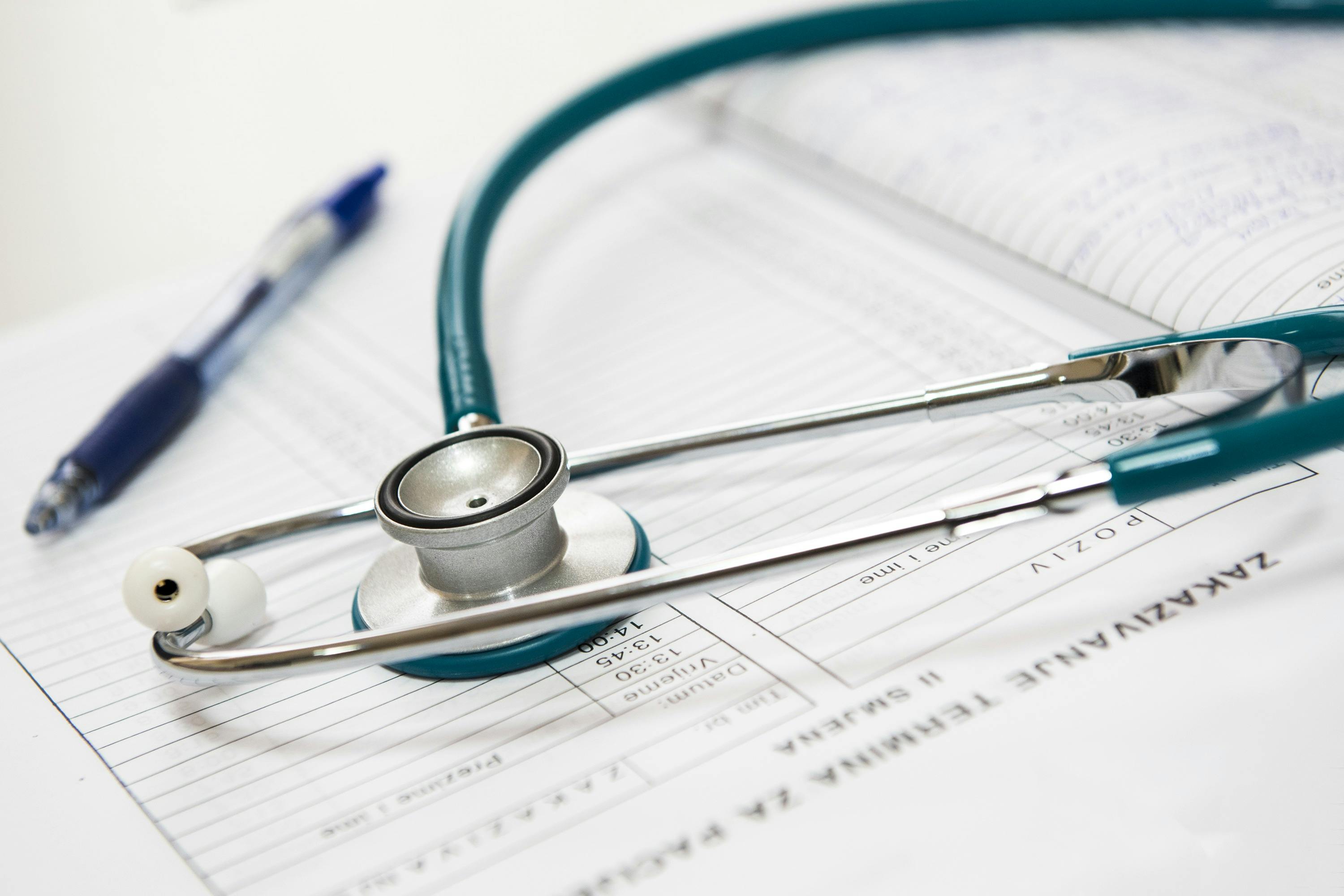
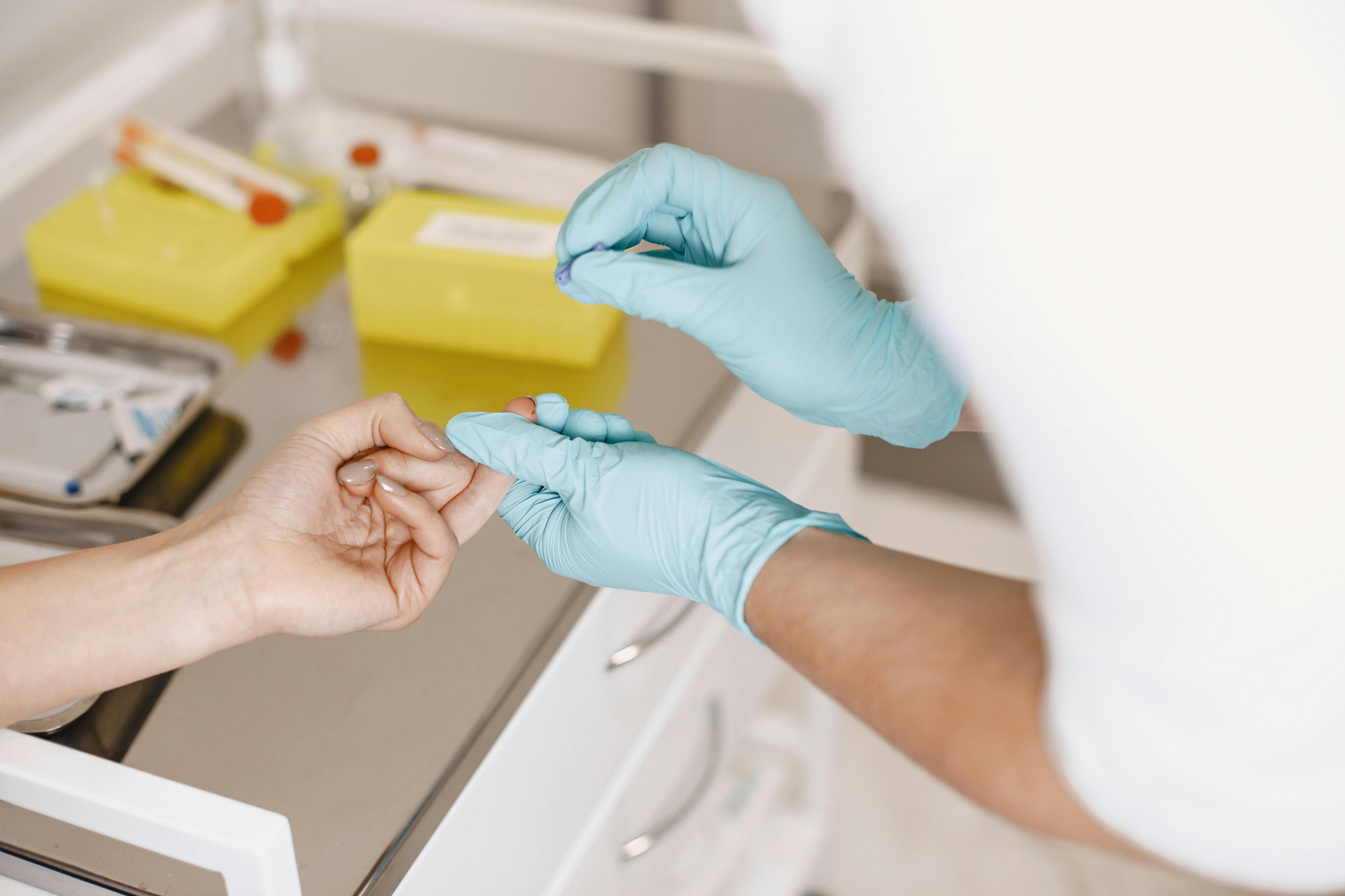
Diagnosis of oral cancer typically involves a visual and physical exam followed by confirmatory tests, such as incisional biopsies, brush biopsies, and imaging tools (e.g., CT or MRI). Biopsies remain the gold standard for diagnosis—but brush biopsy offers a less invasive path for initial screening.
Treatment relies on the cancer’s stage and may include surgery for localized lesions, radiation therapy, or chemotherapy. Recent advances also incorporate targeted molecular therapies and immunotherapy options for certain patient subgroups.
Prognosis and Prevention
Survival rates increase dramatically if cancer is discovered early. Preventive strategies are paramount and include cessation of tobacco, moderated alcohol use, HPV vaccination, and semi-annual oral screenings. New technologies like brush biopsy can also encourage earlier evaluations among asymptomatic patients, particularly in demographics prone to postponing care.
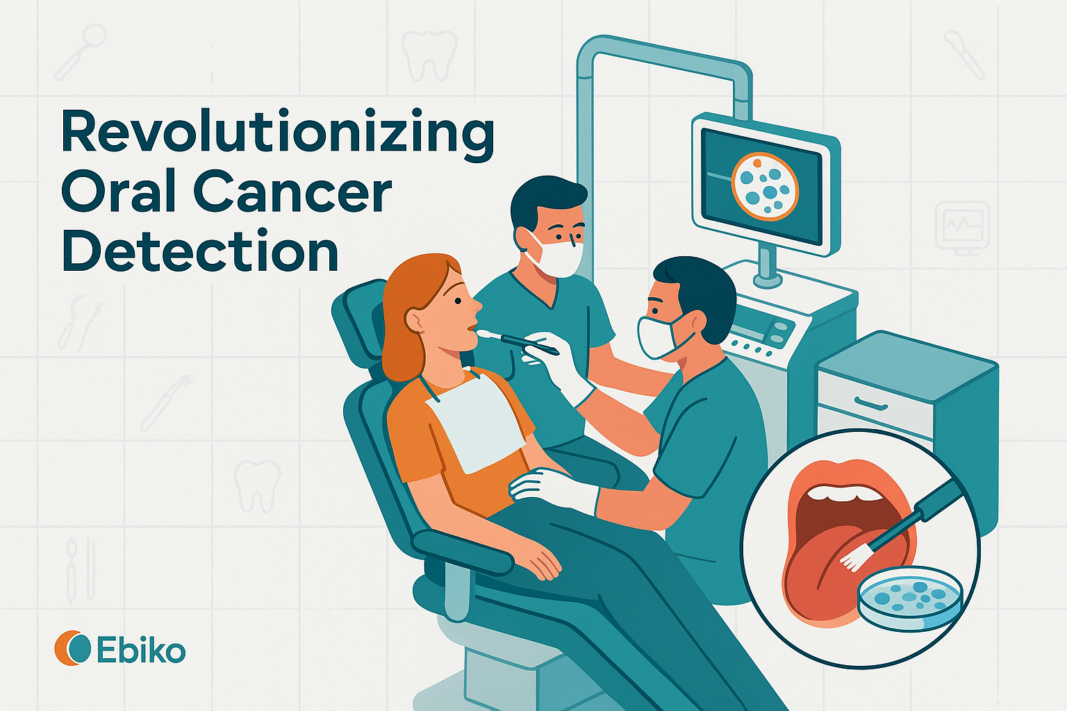
The Breakthrough: Brush Biopsy
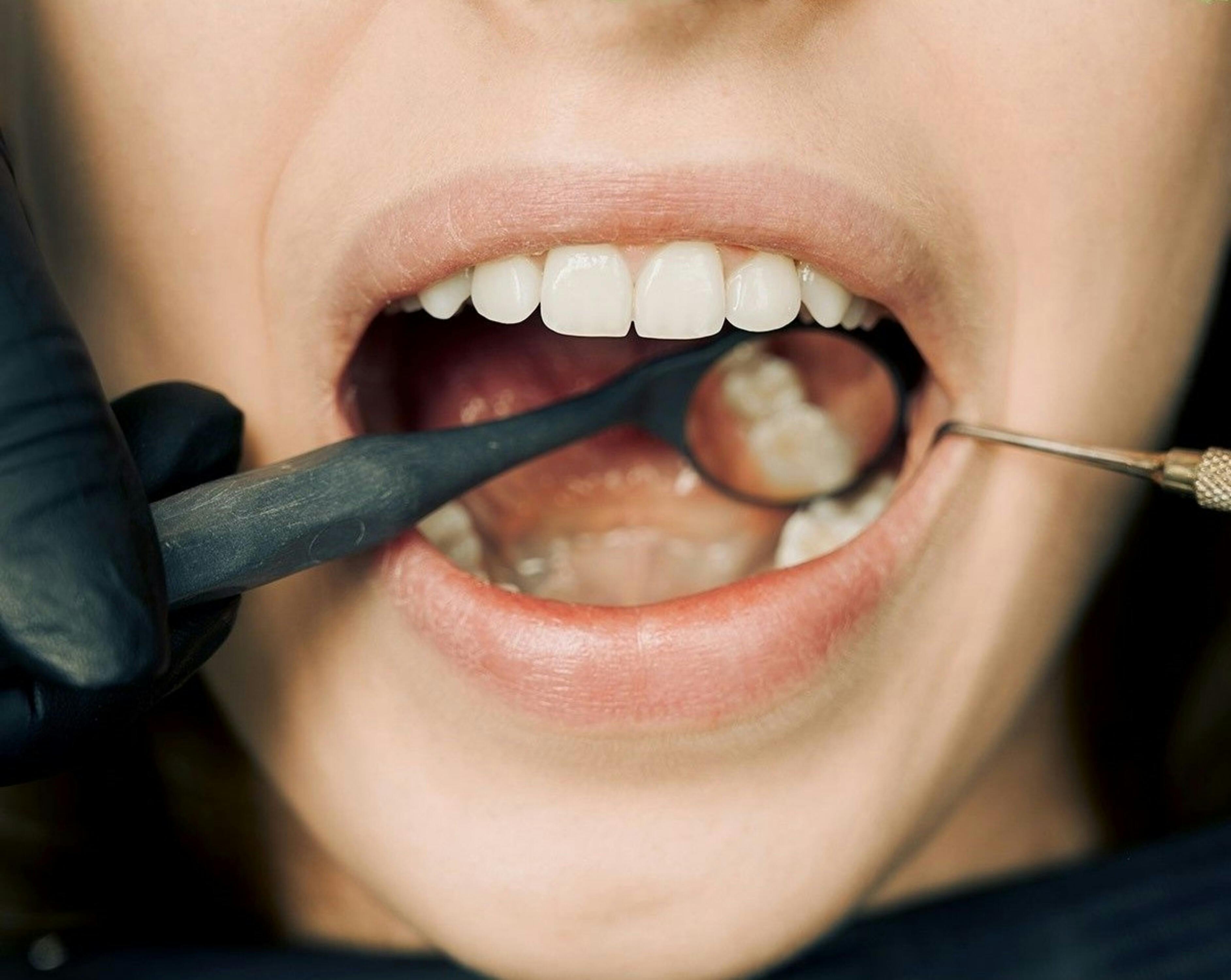
The brush biopsy technique, developed by Guy Adami and Dr. Joel Schwartz, was engineered to make early detection of oral squamous cell carcinoma (OSCC) more accessible. Instead of performing a scalpel biopsy that often requires anesthesia and involves tissue cutting, the brush biopsy uses a small, soft brush to collect a full transepithelial tissue sample.
How Brush Biopsy Works
Clinicians gently press and rotate a sterile brush against the mucosal surface of the oral lesion. The entire epithelium is sampled—from the superficial to the basal layer—without requiring an incision. The collected cells are then fixed and sent for cytological analysis by a pathology laboratory.
Advantages Over Traditional Methods
- Non-invasive and rapid—can be completed chairside
- Much higher patient acceptance due to the absence of pain or bleeding
- Useful for documenting and tracking lesion changes over time
- Low-risk pathway to identify candidates requiring immediate biopsy referral
Future Prospects in Dentistry

The commercialization of brush biopsy tools through companies like Arphion Diagnostics opens the door to advanced diagnostic analytics, such as machine learning and microRNA analysis. This evolution in cytopathology could soon enable dental professionals to not only detect OSCC, but other systemic conditions exhibiting oral manifestations using the same brushing technique.
Integrating Brush Biopsy in Dental Practice

For dental professionals in Toronto and across Canada aiming to elevate their diagnostic capabilities, integrating brush biopsy into routine practice is a logical and impactful step. Here's a pragmatic workflow:
- Identify: During standard exams, flag any lesion not healing after two weeks—for example, unexplained ulcers, pigment changes, or keratoses.
- Perform: Carry out a brush biopsy. No anesthesia is needed, and the total procedure typically takes under five minutes chairside.
- Analyze: Prepare and send the sample to a qualified pathology lab for cytologic interpretation.
- Follow-Up: For negative findings, document and monitor. For positive findings, refer for a confirmatory scalpel biopsy and coordinate with an oral surgeon or oncologist promptly.
EBIKO Dental supplies several resources, such as diagnostic devices and oral screening tools, that pair well with brush biopsy workflows—ensuring clinics have the right tools to deliver proactive care.
Conclusion: The Impact of Brush Biopsy
Oral cancer rates continue to rise in North America, and Toronto—with its large, diverse, and aging population—must rise to the challenge of scalable detection strategies. The brush biopsy gives dental professionals a low-barrier tool for early identification, allowing faster decision-making and improved outcomes.
“By leveraging non-invasive tools like the brush biopsy and combining them with systems like VELScope, dentists in Toronto can significantly improve early detection rates of oral cancer,” says Dr. Perrone, summarizing the larger public health implications.
Brush biopsy is poised to become a cornerstone of contemporary dental diagnostics. Embracing its integration today equips practices with a new standard of care that every patient deserves—and can help save lives across our communities.