TL;DR: A clinical study from the Federal University of Pelotas in Brazil, published online in Clinical Oral Investigations on October 19, 2026, tested whether ordinary 3D intraoral scanner images can detect caries around existing restorations. Across 299 restorations in 41 adults, the scanner was highly specific (84.1% to 99.7%) but missed many lesions, catching only 40% of moderate ones. The authors call it a complementary tool, not a replacement for the visual-tactile exam and radiographs.
Intraoral scanners were initially developed as an alternative to traditional impressions. As their clinical use has increased, researchers have started testing them as diagnostic aids. As of October 2026, a new diagnostic accuracy study asks a practical question for any practice in Toronto or the GTA that already owns a scanner: can ordinary scans help you spot recurrent decay at the margins of existing restorations?
The short answer from this study is "partly." Specificity was high, especially at the moderate and severe thresholds (97.4% to 99.7%), but the scanner under-called lesions in the middle of the severity range. Here is what the researchers did, what they found, and how to read the numbers before changing anything in your recall workflow.
What did the Pelotas study test?
The study was run at the dental clinic of the School of Dentistry, Federal University of Pelotas (UFPel), Brazil, with data collected from July 2023 to September 2024. The authors are Fernandes, Montagner, Karam, van de Sande and Correa. They enrolled adults aged 18 to 60 who had amalgam or composite restorations on anterior or posterior permanent teeth. In total, 299 restorations in 41 patients were assessed: 253 posterior and 46 anterior.
Every restoration was examined two ways:
- Clinical reference standard: after prophylaxis, trained and calibrated examiners used a mirror, a ball-point probe, the operatory light and air drying, scoring each restoration with the CARS criteria.
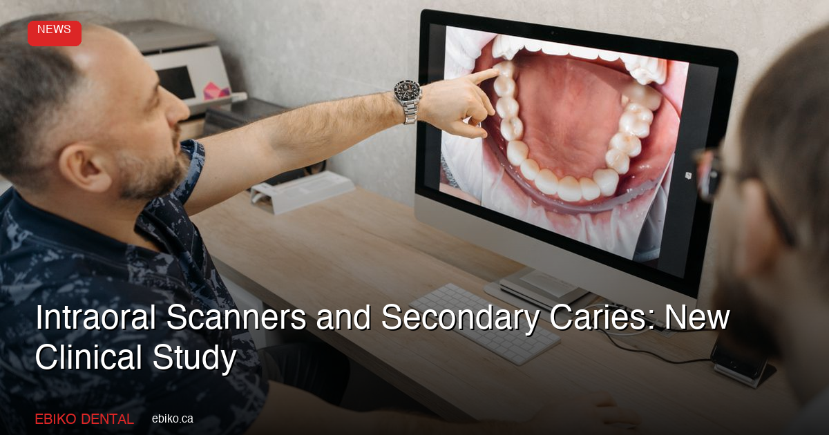
- Index test: the teeth were scanned with a TRIOS 3 scanner (3Shape) under standard white light, kept moist. One week later, a separate calibrated examiner, blinded to the clinical scores, reviewed the 3D models on a high-resolution screen.
CARS stands for Caries Associated with Restorations or Sealants. The authors describe it as a 0 to 6 scale from the International Caries Classification and Management System (ICCMS), where scores of 4 or higher generally indicate operative intervention. Because some CARS features depend on air-drying, wet-surface observation or probing, which a static scan cannot reproduce, the image findings were grouped into four categories: sound, initial, moderate and severe.
The authors state that, to their knowledge, no earlier clinical validation study had tested conventional scanner images for caries around restorations using CARS. Most prior work looked at primary caries on unrestored teeth or at scanners with add-on optical caries-detection features.
How accurate was the scanner?
The researchers measured performance at three cumulative thresholds: any lesion (initial and above), moderate and above, and severe only.
The headline figures, as reported by the authors:
- Sensitivity: 75% at the initial threshold, 40% at the moderate threshold and 57.1% at the severe threshold.
- Specificity: 84.1%, 97.4% and 99.7% at the same three thresholds.
- Positive predictive value: 60%, 58.8% and 80%. Negative predictive value: 91.4%, 94.7% and 99.0%.
- Agreement with the clinical exam: exact agreement of 77.9%, linearly weighted agreement of 91.1%, and a weighted kappa of 0.53, which the authors describe as moderate.
The study also reports "AUC" values of 0.79, 0.69 and 0.78. Read these carefully: because the scanner calls were binary, the authors calculated AUC as the average of sensitivity and specificity (balanced accuracy), not from a full ROC curve.
Why did moderate lesions slip through?
Moderate lesions were the weak spot, with sensitivity of 40%. The authors explain that this category includes small enamel cavities and dentin shadowing. A ball-point probe can detect those small cavities chairside; on a screen, where the assessment is visual only, some of them go unseen.
The authors add that because CARS was adapted to images, sensitivity at the moderate threshold "may have been underestimated," and that radiographs may help resolve uncertainty for moderate lesions. In EBIKO Dental's reading, a tool that misses a share of moderate lesions can support documentation, but it should not be the deciding exam.
The severe-lesion numbers need a different caution. Only 7 of the 299 restorations (2.3%) were clinically severe. The scanner classified 4 of those 7 as severe and placed the other 3 in lower categories. With so few cases, the confidence interval for severe-threshold sensitivity runs from 33.3% to 100%, and the authors say a larger sample would be needed to pin it down.
Pro Tip: The study's authors note that scanner images may help document and assess visible changes around restorations. Our suggestion: if your team already scans, save each scan to the chart with its date so the same restoration can be compared across visits.
What are the study's limits?
The authors list several limitations, and we add two observations of our own at the end:
- An imperfect reference standard. The comparator was an expert visual-tactile exam, not histology. The authors acknowledge this standard does not remove diagnostic uncertainty.
- No repeat readings. Each restoration was scored clinically by a single examiner, and the scan images were read once, so intra-examiner reproducibility for the scan reading was not estimated. Before the study, the image examiner reached 94.8% and 90.1% agreement (weighted kappa 0.80 and 0.75) with the two clinical examiners during calibration.
- An adapted scale. CARS was designed for direct clinical examination. The authors call their on-screen use "an image-based adaptation" and recommend developing a scanner-specific version.
- No radiographs in the reference standard. The authors list the absence of radiographs as part of the reference standard as a limitation.
- Cost. The authors note the high cost of intraoral scanners may limit their applicability in some clinical settings.
- Our observation: one scanner, white light only. The study used a TRIOS 3 under standard white light. It did not test the fluorescence or near-infrared caries features offered on some scanners, which have been examined in other studies.
- Our observation: one university clinic. Results come from 41 patients at one Brazilian dental school.
What does this mean for Canadian dental practices?
The authors' conclusion is measured: the scanner is "a complementary tool," and clinical interpretation should include visual-tactile examination and radiographic assessment when indicated. The study did not look at Canadian practices. Our reading, for practices in Toronto, Mississauga, Markham and across Ontario that already own a scanner:
- Treat a negative scan as reassuring, not conclusive. Negative predictive values were 91.4% to 99.0% in this study, but a clean-looking scan did not rule out moderate lesions.
- Keep the probe and the radiograph in the workflow. The features the scan could not reproduce (air-drying response, wet-surface appearance, tactile confirmation) are exactly the ones the authors identified as limits.
- Use scans for what the authors say they can do. Their clinical-relevance statement supports using scanner images to document and assess visible changes around restorations.
The authors also note that, alongside fractures, secondary caries is a leading reason restorations are replaced. They chose the CARS criteria for its more conservative, less invasive approach, noting that the FDI criteria tend to be more interventionist because of their focus on marginal defects.
Pro Tip: Our suggestion: when a scan suggests marginal change but the clinical exam is equivocal, record both findings and set a re-check date rather than replacing the restoration on the image alone.
EBIKO Dental will continue monitoring research on digital diagnostics and what it means for Canadian practices. For clinical supplies and practice essentials, visit ebiko.ca.
Sources
- Fernandes BM, Montagner AF, Karam SA, van de Sande FH, Correa MB. Validation of 3D intraoral scanner use for detecting caries lesions around dental restorations: A diagnostic accuracy study. Clinical Oral Investigations. 2026;30(11). doi:10.1007/s00784-026-07226-7 (open-access full text: Europe PMC)
Frequently Asked Questions
Q: Can an intraoral scanner detect caries around existing fillings?
Partly. In a 2026 study of 299 restorations, standard white-light scanner images caught 75% of lesions at any severity but only 40% of moderate-or-worse lesions, with specificity of 84.1% to 99.7%. The authors recommend using scans alongside the visual-tactile exam and radiographs, not instead of them.
Q: What are the CARS criteria?
CARS (Caries Associated with Restorations or Sealants) is a 0 to 6 visual scoring system from the International Caries Classification and Management System. According to the study's authors, scores of 4 or higher generally indicate operative intervention, with decisions also weighing caries risk and other findings.
Q: Did the study test scanner caries-detection features like fluorescence?
No. The researchers used a TRIOS 3 scanner under standard white light only. Scanners with fluorescence or near-infrared features were not evaluated in this study.